We utilise iPSC microglia as In vitro models for microglia mediated neuroinflammation. iPSC Microglia were characterised for TMEM119 and CD11b phenotypic markers (by cell imaging and flowcyte) and validated by cytokine release, phagocytosis, chemotaxis, and cell morphology.
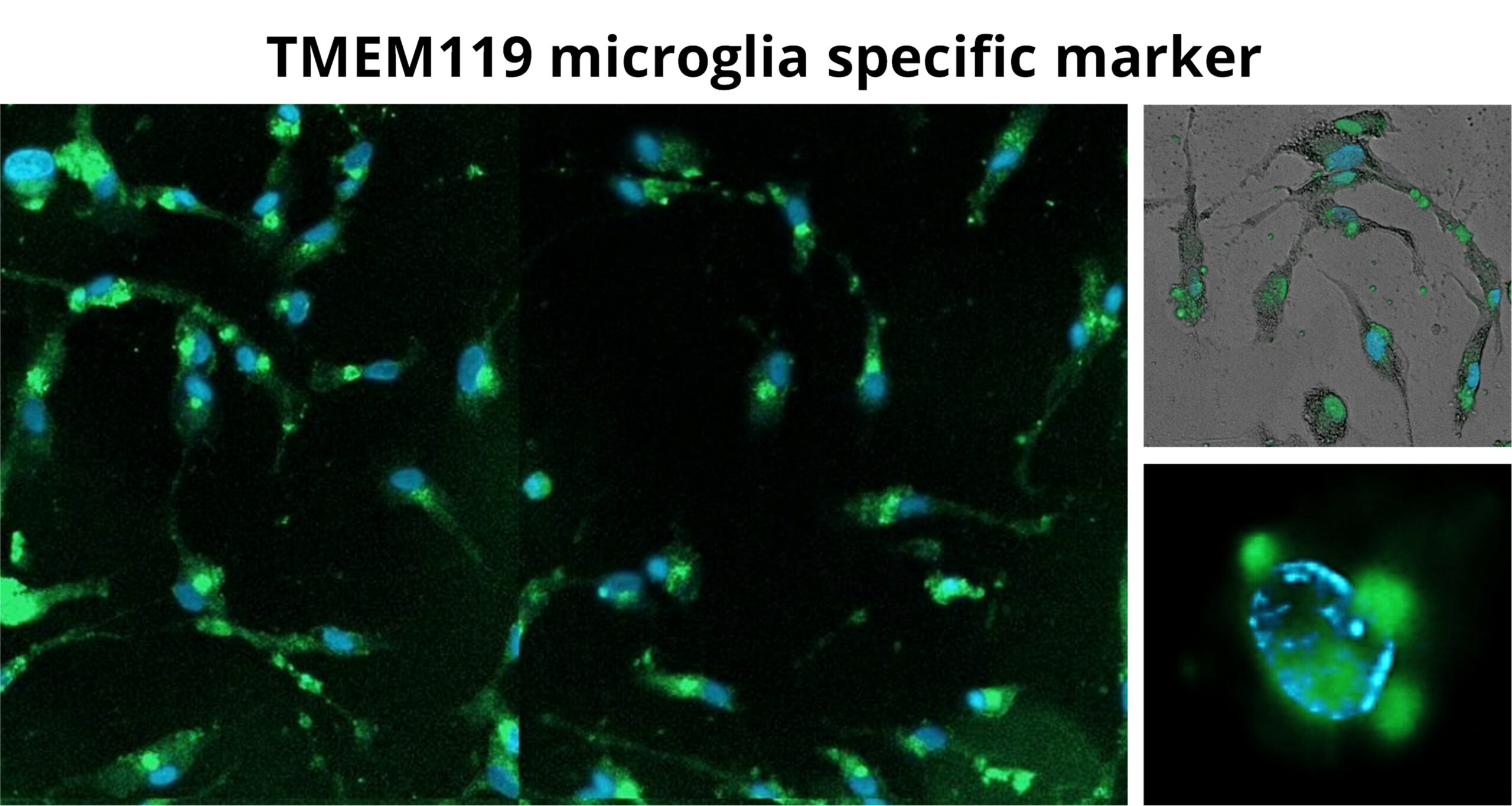
Figure-A: iPSC-derived microglia were matured for 8 days. Microglia stained with Blue Nuclear marker, and Green TMEM119 microglia specific marker, image taken using ImageXpress pico.
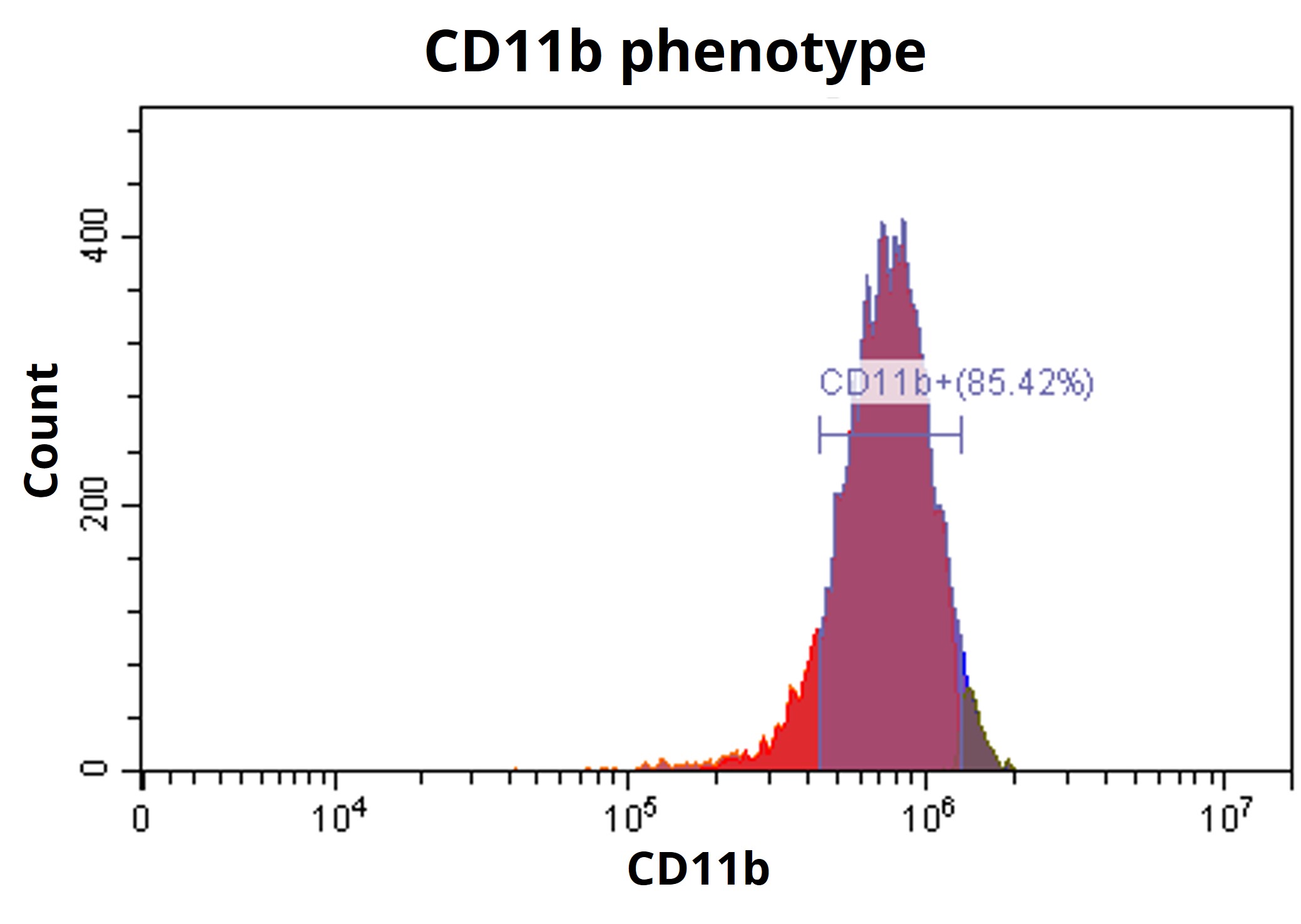
Figure-B: Microglia expressing CD11b phenotypic marker measured by Flowcytometry.
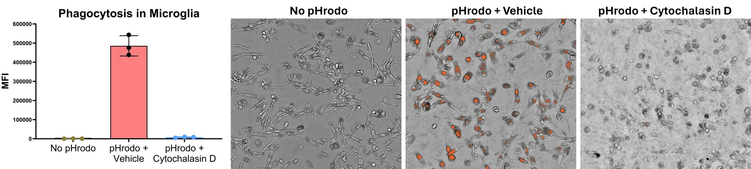
Figure-C: Phagocytosis. Matured iPSC microglia were labeled with pHrodo, and phagocytosis was measured after 24 hours in the presence and absence of Cytochalasin-D (10uM). Cytochalasin-D showed complete inhibition of phagocytosis.
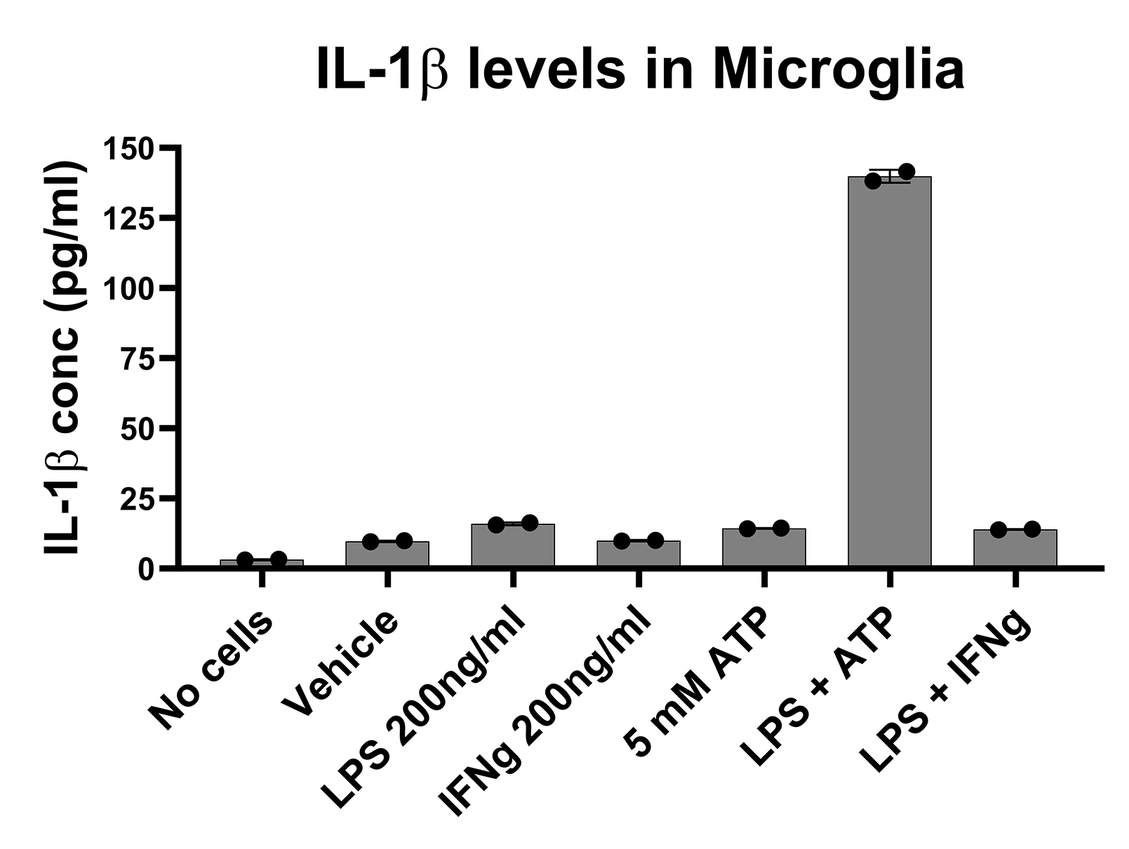
Figure-D: IL-1 secretion from matured iPSC microglia.
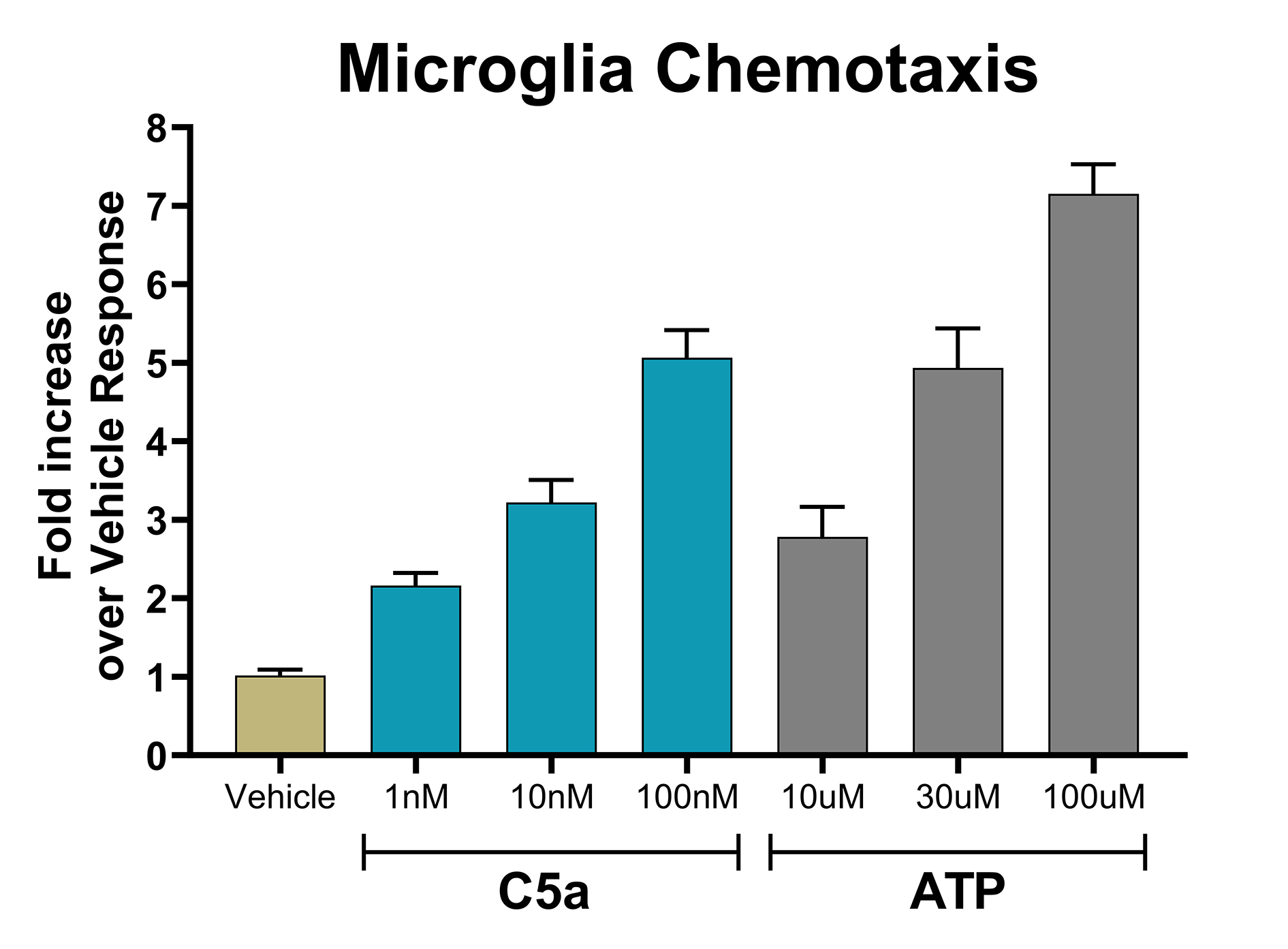
Figure-E: Fold increase in Microglia migration upon addition of C5a & ATP.
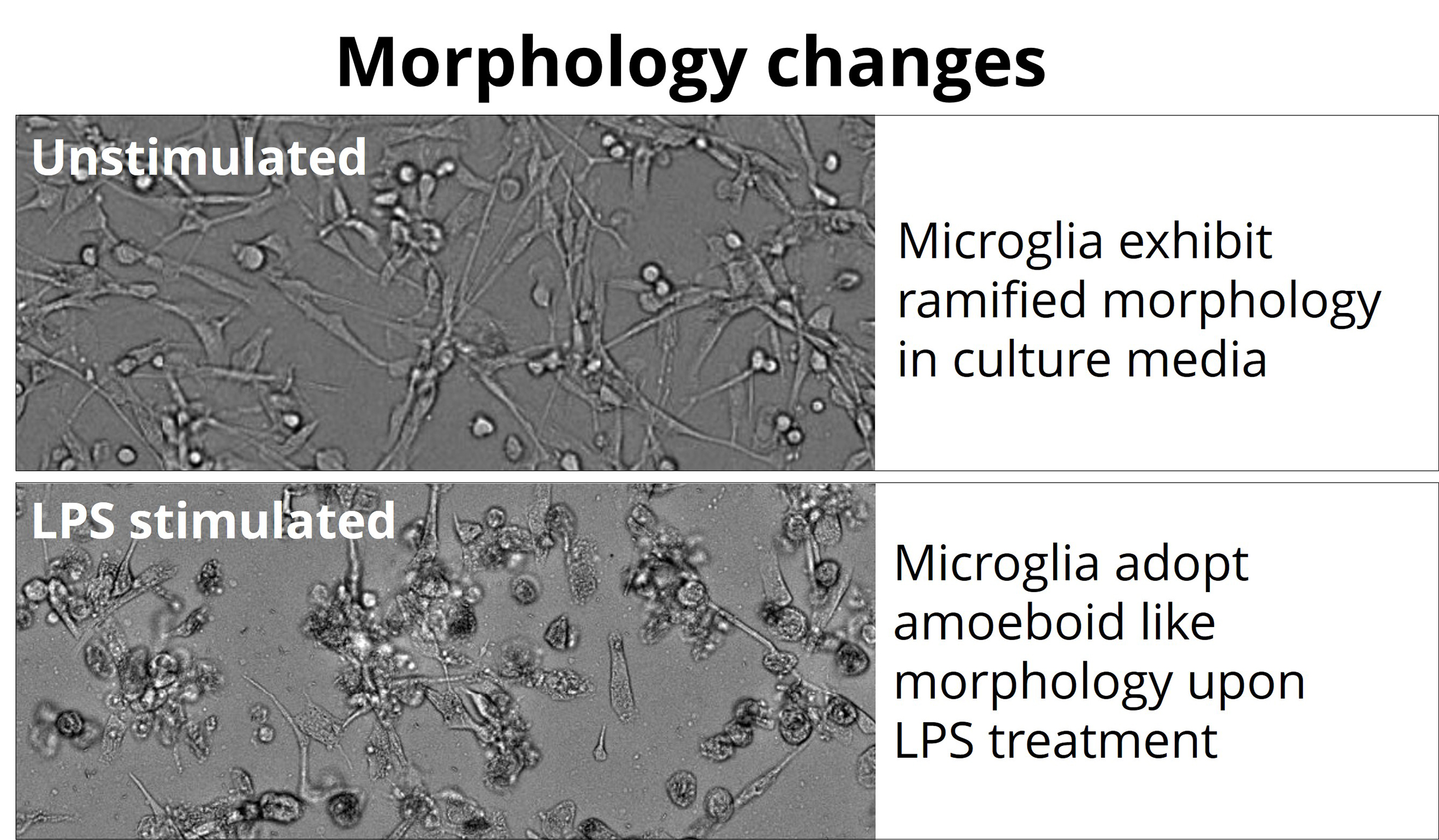
Figure-F: Microglia morphology check brightfield images.
Here we demonstrate successful culture of iPSC derived microglia, characterised by expression of microglia phenotypic markers, and validated by functional response pattern demonstrating relevance. Validated microglia assays at BioMedha enabled us supporting client’s programmes, measuring compound effect (to assess therapeutic efficacy) on Microglia function and gene expression.
© Copyright BioMedha Limited. Company Reg no: 09872164 | VAT reg no: GB 303220569
© Copyright BioMedha Limited
| Cookie | Duration | Description |
|---|---|---|
| cookielawinfo-checkbox-analytics | 11 months | This cookie is set by GDPR Cookie Consent plugin. The cookie is used to store the user consent for the cookies in the category "Analytics". |
| cookielawinfo-checkbox-functional | 11 months | The cookie is set by GDPR cookie consent to record the user consent for the cookies in the category "Functional". |
| cookielawinfo-checkbox-necessary | 11 months | This cookie is set by GDPR Cookie Consent plugin. The cookies is used to store the user consent for the cookies in the category "Necessary". |
| cookielawinfo-checkbox-others | 11 months | This cookie is set by GDPR Cookie Consent plugin. The cookie is used to store the user consent for the cookies in the category "Other. |
| cookielawinfo-checkbox-performance | 11 months | This cookie is set by GDPR Cookie Consent plugin. The cookie is used to store the user consent for the cookies in the category "Performance". |
| viewed_cookie_policy | 11 months | The cookie is set by the GDPR Cookie Consent plugin and is used to store whether or not user has consented to the use of cookies. It does not store any personal data. |